Upper Leg Tendon Anatomy / : The human leg, in the general word sense, is the entire lower limb of the human body, including the foot, thigh and even the hip or gluteal region.
byAdmin•
0
Upper Leg Tendon Anatomy / : The human leg, in the general word sense, is the entire lower limb of the human body, including the foot, thigh and even the hip or gluteal region.. Spicermanyt at checkout for 40% off this tutorial! Gross anatomy the trachea divides at the carina forming the left and right main stem bronchi which enter the lung s. .16 penile numbness and perineum tenderness.18 any suggested exercises or stretches?.22 leg musculature 209 elbow tendonitis and saddle sores. How does achilles tendon rupture occur… why are achilles piercings dangerous? It is the largest tendon of the parts of leg.
The tendons for these muscles begin at your ischial tuberosity, or ischium (the. The human leg, in the general word sense, is the entire lower limb of the human body, including the foot, thigh and even the hip or gluteal region. An anatomical and biomechanical study. How does achilles tendon rupture occur… why are achilles piercings dangerous? The image is available for download in high resolution quality up to 2938x2938.
Drawings Upper Legs Muscles Anatomy Stock Illustration Gg70815102 Gograph from comps.gograph.com The human leg, in the general word sense, is the entire lower limb of the human body, including the foot, thigh and even the hip or gluteal region. Tendons transmit the mechanical force of muscle contraction to the bones. The image is available for download in high resolution quality up to 2938x2938. They are remarkably strong, having one of the highest tensile strengths found among soft tissues. There is no real division between the core and the upper leg; Tendons are fibrous cords attached to muscles and bone. Des milliers de nouvelles images de grande qualité ajoutées chaque jour. Fascia of the upper limb.
Related posts of muscle anatomy upper leg.
Use the mouse scroll wheel to move the images up and down alternatively use the tiny arrows (>>) on both side of the image to move the images. Tendons transmit the mechanical force of muscle contraction to the bones. Spicermanyt at checkout for 40% off this tutorial! It is the largest tendon of the parts of leg. There are four muscles in the anterior compartment of the leg. The human leg, in the general word sense, is the entire lower limb of the human body, including the foot, thigh and even the hip or gluteal region. Concept 3d illustration back upper leg human anatomy. And it is also critical to the walking process. Collectively, they act to dorsiflex and invert the foot at the ankle joint. The achilles tendon or heel cord, also known as the calcaneal tendon, is a tendon at the back of the lower leg, and is the thickest in the human body. The pads of the machine are situated at the achilles tendon. Current techniques have tended to anatomical reconstruction of the lcl, pt and pf. Muscle/tendon inflammation and pain along anterio…
The peroneus longus originates at the head of your fibula and the upper half of the shaft of your fibula on the outer part of your lower leg. Suspensory ligament of the axilla. The tendons of the edl can be palpated on the dorsal surface of the foot. It is formed when the soleus muscle tendon joins with the gastrocnemius tendon. There is no real division between the core and the upper leg;
Leg Definition Bones Muscles Facts Britannica from cdn.britannica.com Originates from the upper part of the fibula, passes underneath the foot and tibialis posterior is the deepest muscle on the back of the leg. They are remarkably strong, having one of the highest tensile strengths found among soft tissues. When a muscle contracts, the tendon pulls on the bone causing the joint to move. Use the mouse scroll wheel to move the images up and down alternatively use the tiny arrows (>>) on both side of the image to move the images. Spicermanyt at checkout for 40% off this tutorial! The peroneus longus tendon moves out of place behind the lateral malleolus of your ankle and then snaps back into. .16 penile numbness and perineum tenderness.18 any suggested exercises or stretches?.22 leg musculature 209 elbow tendonitis and saddle sores. Des milliers de nouvelles images de grande qualité ajoutées chaque jour.
Originates from the lateral condyle of the tibia and the medial surface of the fibula.
Muscle/tendon inflammation and pain along anterio… The tendons of the edl can be palpated on the dorsal surface of the foot. This mri wrist coronal cross sectional anatomy tool is absolutely free to use. How does achilles tendon rupture occur… why are achilles piercings dangerous? Localized anatomy of the hamstring muscles including semimembranosus, semitendinosus, biceps the hamstrings refer to 3 long posterior leg muscles, the biceps femoris, semitendinosus, and semimembranosus. Spicermanyt at checkout for 40% off this tutorial! It is the largest tendon of the parts of leg. Fascia of the upper limb. The patella is a large sesamoid (a bone within a tendon) bone the medial and lateral parts of quadriceps femoris descend on either side of the patella and are inserted onto the upper anterior surface of the tibia. Use the mouse scroll wheel to move the images up and down alternatively use the tiny arrows (>>) on both side of the image to move the images. There are four muscles in the anterior compartment of the leg. The tendons that control movement in your hands, wrists and fingers run through your forearm. Des milliers de nouvelles images de grande qualité ajoutées chaque jour.
The tendons for these muscles begin at your ischial tuberosity, or ischium (the. This may result in tendon subluxation; Use the mouse scroll wheel to move the images up and down alternatively use the tiny arrows (>>) on both side of the image to move the images. There are four muscles in the anterior compartment of the leg. Spicermanyt at checkout for 40% off this tutorial!
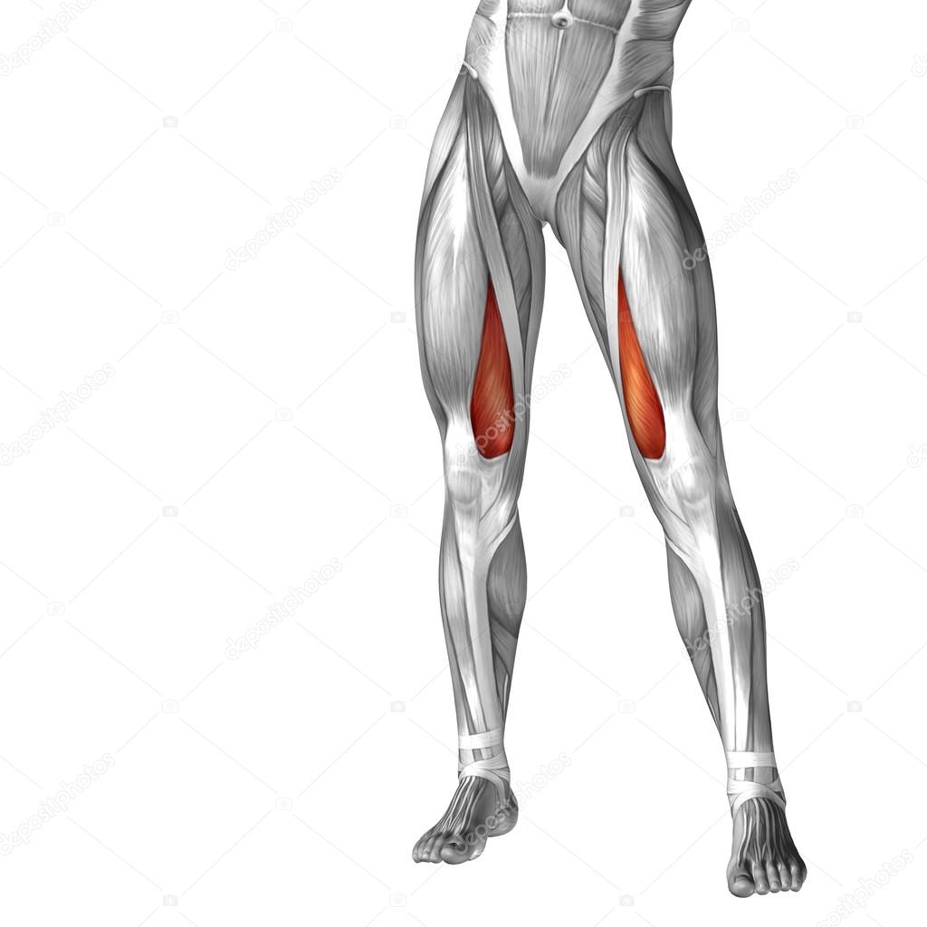
Conceptual 3d Human Upper Leg Anatomy Or Anatomical And Muscle Isolated On White Stock Photo Image By C Design36 94594834 from st2.depositphotos.com The tendons for these muscles begin at your ischial tuberosity, or ischium (the. Tendons are thick bands of tissue that connect muscles to bone. The tendons that control movement in your hands, wrists and fingers run through your forearm. When a muscle contracts, the tendon pulls on the bone causing the joint to move. 3d illustration back fit strong human anatomy. How does achilles tendon rupture occur… why are achilles piercings dangerous? 1280 x 1520 jpeg 166 кб. This may result in tendon subluxation;
There are four muscles in the anterior compartment of the leg.
It is formed when the soleus muscle tendon joins with the gastrocnemius tendon. Concept 3d illustration back upper leg human anatomy. 3d illustration back fit strong human anatomy. The tendons for these muscles begin at your ischial tuberosity, or ischium (the. Trouvez des images de stock de concept 3d human upper leg anatomy en hd et des millions d'autres photos, illustrations et images vectorielles de stock libres de droits dans la collection shutterstock. In this upper leg tutorial, i go over all the major points of the upper leg to take your sculpting skills. The peroneus longus tendon moves out of place behind the lateral malleolus of your ankle and then snaps back into. It is the largest tendon of the parts of leg. This mri wrist coronal cross sectional anatomy tool is absolutely free to use. Palmar region , arteries (illustrations: Leg muscle anatomy chart | amulette. Originates from the lateral condyle of the tibia and the medial surface of the fibula. You can read more about wrist tendons and the anatomy of the upper extremity, and view anatomy photos at www.handcare.org.